Fig. 1.
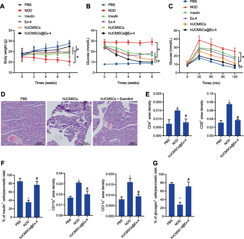
hUCMSCs@Ex-4 promotes the repair of islet tissue damage by reducing the blood glucose level in NOD mice. A Body weight of NOD mice after different treatments. B Blood glucose level in NOD mice with different treatments. C Blood glucose level in NOD mice with different treatments measured by OGTT. D HE staining for islet morphology in NOD mice with different treatments (400 ×). E Immunohistochemical staining analysis of the infiltration of immune cells (CD4+ T, CD8+ T, CD11b+, and CD11c+) in the pancreatic tissues of NOD mice with different treatments. F Proportion of β-cells in the pancreatic tissues of NOD mice with different treatments analyzed by immunohistochemical staining. G Proportion of α-cells in the pancreatic tissues of NOD mice with different treatments analyzed by immunohistochemical staining. In panels A-C, *p < 0.05 vs. NOD mice. #p < 0.05, NOD mice treated with hUCMSCs@Ex-4 vs. NOD mice treated with hUCMSCs. In panels E–G, *p < 0.05 vs. PBS-treated mice. #p < 0.05 vs. NOD mice. n = 12

