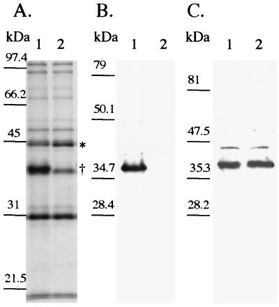
FIG. 3.
(A) SDS–10% PAGE and Coomassie blue staining of OMP prepared from 35000HP (lane 1) and 35000HP-SMS2 (lane 2). Note the location of the 45-kDa protein (∗) and the absence of MOMP (†). (B and C) Western blot analysis of 35000HP (lane 1) and 35000HP-SMS2 (lane 2). Panel B was probed with MAb 3F12, and panel C was probed with MAb 2C7. Molecular mass standards are shown to the left of each panel.

