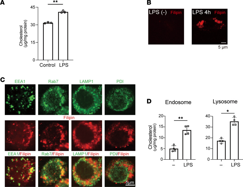
Figure 1. Cellular cholesterol accumulates in response to TLR4 activation in RAW cells.
RAW cells cultured in medium containing 10% FBS were stimulated with or without LPS (100 ng/mL) for 4 hours. (A) Cellular unesterified free cholesterol was quantified using gas chromatography/mass spectrometry (GC/MS). n = 4 in each group. **P < 0.01, Student’s 2-tailed t test. (B) Cellular free cholesterol was stained with filipin. Scale bar, 5 μm. (C) Localization of filipin staining within early and late endosomes and lysosomes. RAW cells treated for 4 hours with LPS were stained with markers for organelles [EEA1 (early endosomes), Rab7 (late endosome), LAMP1 (lysosomes), and PDI (ER)] (green) and with filipin (red). Scale bar, 5 μm. (D) Cholesterol levels within endosomes and lysosomes were measured using GC/MS. n = 4 in each group. *P < 0.05, **P < 0.01, Student’s 2-tailed t test. Data shown as mean ± SD in all panels where P values are shown.

