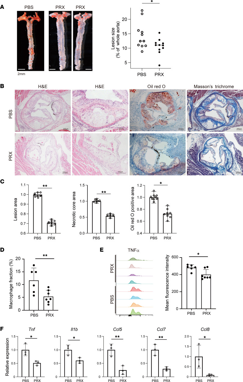
Figure 9. PRX suppresses atherogenesis in Ldlr–/– mice.
(A) Male Ldlr–/– mice were fed a high-cholesterol diet for 11 weeks, with or without subcutaneous PRX injection (1,000 mg/kg BW every 2 days). Aortic atheromatous lesions were visualized after staining with Oil Red O. Oil Red O–positive areas were quantified and normalized with the entire aortic area. n = 11 in each group. *P < 0.05. Student’s 2-tailed t test. (B) Representative photographs of aortic sinuses from Ldlr–/– mice treated with PBS or PRX. Sections were analyzed by staining with hematoxylin-eosin (H&E), Oil Red O, and Masson’s trichrome. Scale bars, 200 μm or 100 μm (second H&E column). (C) Atherosclerotic lesion, necrotic core, and Oil Red O–positive areas were quantified and normalized to the control values (PBS). n = 10 in each group. **P < 0.01, *P < 0.05. Student’s 2-tailed t test. (D) Flow cytometric analysis of cells from thoracic aortas of mice treated with PRX or PBS. Shown are fractions of CD11b+Ly6G–F4/80+ macrophages among the total live cells. n = 6–7 mice for each group. **P < 0.01, Student’s 2-tailed t test. (E) TNF-α expression in CD11b+Ly6G–F4/80+ macrophages. Note that there were fewer cells expressing higher levels of TNF-α in the PRX-treated group. n = 4 mice for each group. *P < 0.05, Student’s 2-tailed t test. (F) Relative mRNA expression of proinflammatory genes in the aorta. *P < 0.05, **P < 0.01, Student’s 2-tailed t test. Data shown as mean ± SD in all panels where P values are shown.

