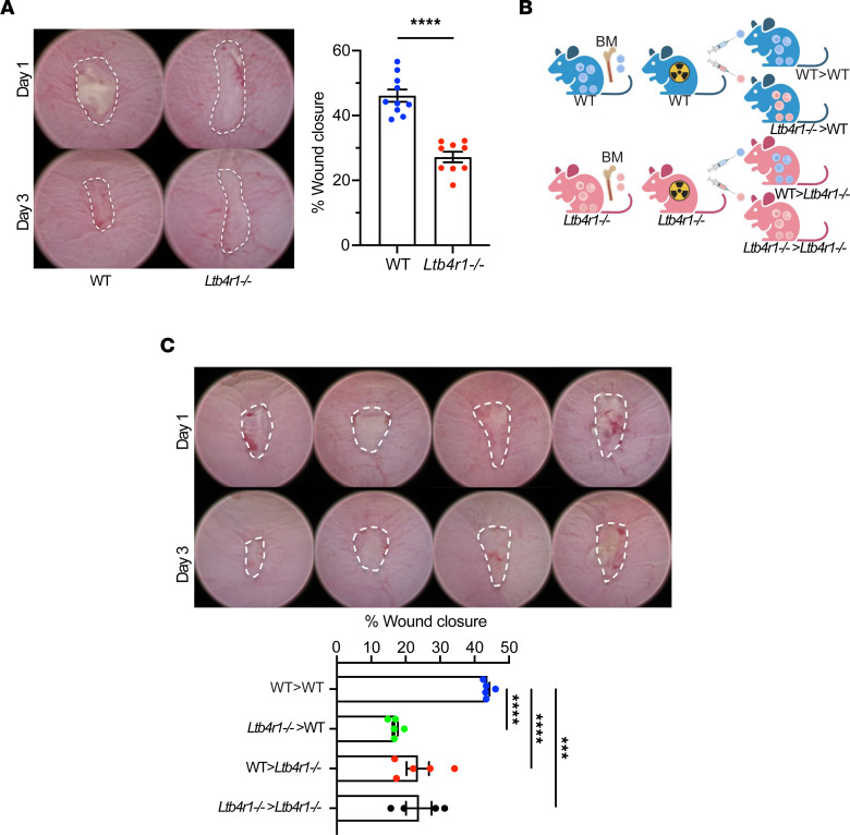
Figure 5. Role of BLT1 in intestinal mucosal wound repair in vivo.
(A) In vivo intestinal mucosal wound repair in Ltb4r1–/– mice. Utilizing a miniature video endoscope and biopsy scissors, 5 wounds were created in the dorsal aspect of the colonic mucosa of anesthetized mice. Digital images of wound surface area at 1 and 3 days after wounding are shown (left). Points represent the mean value within all wounds from individual mice (right). The data are presented as the mean ± SEM of 9 to 10 mice. Statistical analysis was performed using an unpaired (2-tailed) t test with Welch’s correction. ****P < 0.0001. (B and C) In vivo intestinal mucosal wound repair in BM chimeric mice. (B) Illustration of BM chimera experiment. (C) Digital images of wound surface area at 1 and 3 days after wounding are shown (left). Points represent the mean value within all wounds from individual mice (right). The data are presented as the mean ± SEM of 5 mice. Statistical analysis was performed using 1-way ANOVA followed by post hoc Welch’s t test with Bonferroni’s correction. ***P < 0.001, ****P < 0.0001.

