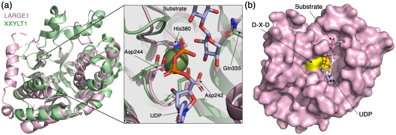
Fig 4. The Xyl-T domain of LARGE1.
(a) Superimposition of the Xyl-T domain from LARGE1 (pink) with XXYLT1 (PDB: 4wn2, green). Inset shows a closeup view of the active site. The conserved residues that coordinate the Mn2+ are noted, as well as a UDP and a growing substrate that were resolved in the structure of XXYLT1. (b) Surface representation of the LARGE1 Xyl-T domain. The aspartic acids of the DXD motif are colored yellow. The UDP and the growing substrate derived from the structure of XXYLT1 are shown as sticks.

