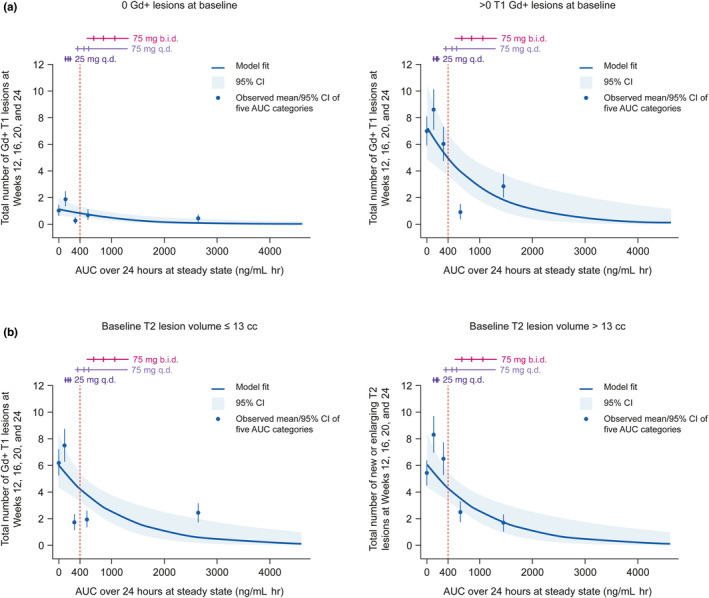
FIGURE 2.

Relationship between (a) total T1 Gd+ and (b) total new/enlarging T2 lesion count and evobrutinib exposure (mITT population). Horizontal lines correspond to the middle 80% of the distribution of AUC by dose/regimen (not split by baseline T1 Gd+ lesion covariate), the three ticks are the first, second (median), and third quartiles. The vertical, dashed lines correspond to an evobrutinib exposure of 400 ng/mL h. Observed means (blue points) are plotted at the mid‐point of the corresponding AUC exposure group. In the T1 Gd+ lesion analysis, data shown correspond to n = 123 with 0 T1 Gd+ lesions at baseline and n = 84 with >0 T1 Gd+ lesions at baseline. In the new/enlarging T2 lesion analysis, data shown correspond to n = 108 with ≤13 cc T2 lesion volume at baseline and n = 99 with >13 cc T2 lesion volume at baseline. AUC, area under the curve; b.i.d., twice daily CI, confidence interval; Gd+, gadolinium‐enhancing; mITT, modified intent‐to‐treat; q.d., once daily.
