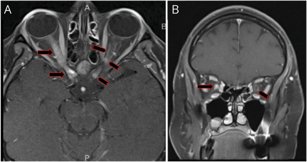
Figure 2. T1, Fat-Suppressed, Postcontrast MRI of the Orbits.
Axial (A) image demonstrates longitudinally extensive enhancement of the right optic nerve extending into the prechiasmatic area (black and red arrow) along with faint contrast enhancement of the left optic nerve (black and red chevron) in a pattern of subtle and very thin ‘tram tracking’, signifying optic nerve sheath enhancement (i.e., perineuritis). Furthermore, there are nodular enhancements involving the left optic nerve sheath at the orbital apex, but also the intracanalicular, prechiasmatic portion of the optic nerve, with very subtle ‘tram tracking’ (black and red chevrons). Coronal image (B) shows diffuse right optic nerve enhancement (black and red arrow) and perineural enhancement of the left optic nerve (black and red chevron), in keeping with the ‘donut sign’ of perineuritis.

