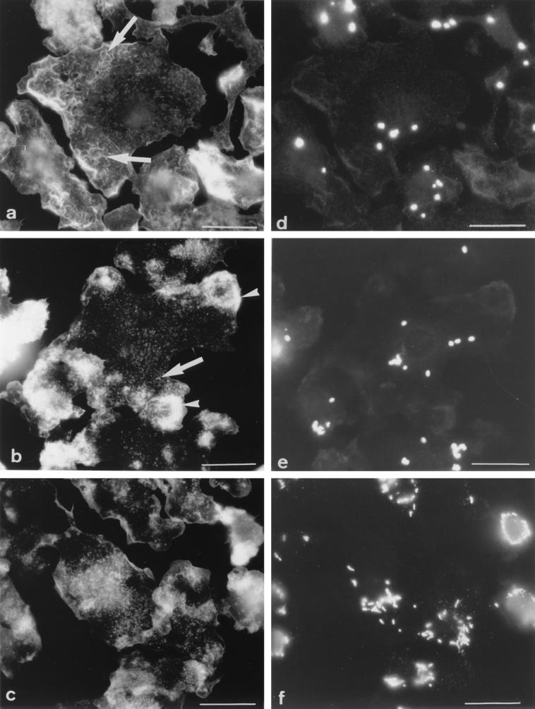
FIG. 2.
Appearance of F-actin in Mφs infected with M. avium. Cells were double labeled for F-actin (a to c) and for immunodetection of bacteria (d to f) at 0 (a and d), 1 (b and e), or 6 (c and f) days after a 4-h infection with M. avium. (a) Day 0: same F-actin pattern as in uninfected cells, i.e., abundance of small filaments (arrows); (b) day 1: accumulation of F-actin in several large patches (arrowhead); (c) day 6: F-actin showing a punctate appearance. In the same cells, phagosomes were scattered at days 0 (d) and 1 (e) and were gathered around the nucleus at day 6 (f). Bars = 5 μm.

