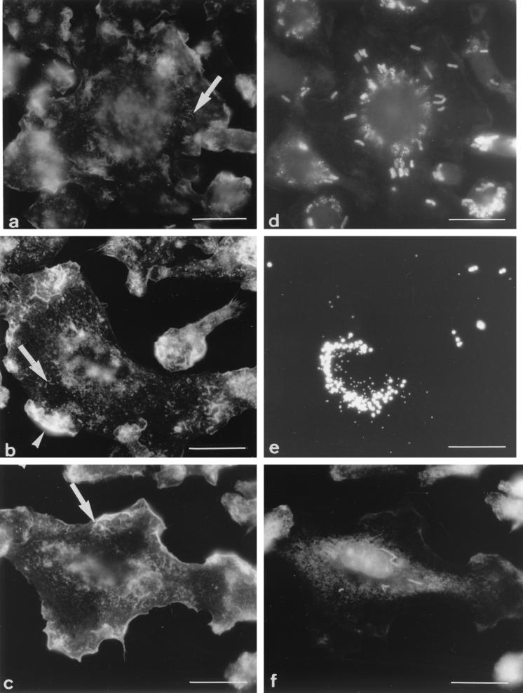
FIG. 3.
Appearance of F-actin (a to c) and location of phagosomes (d to f) after phagocytic uptake of different particles. (a and d) B. subtilis with a 45-min chase after uptake; (b and e) fluoresbrite microspheres with a 2-h chase after uptake; (c and f) M. smegmatis 1 day after phagocytic uptake. In all cases, the F-actin network retained the appearance and distribution pattern observed in uninfected cells, i.e., small filaments (arrows) and zero, one, or two patches (arrowheads). At these time points, phagosomes were gathered around the nucleus in all cases. Bars = 5 μm.

