Figure 1.
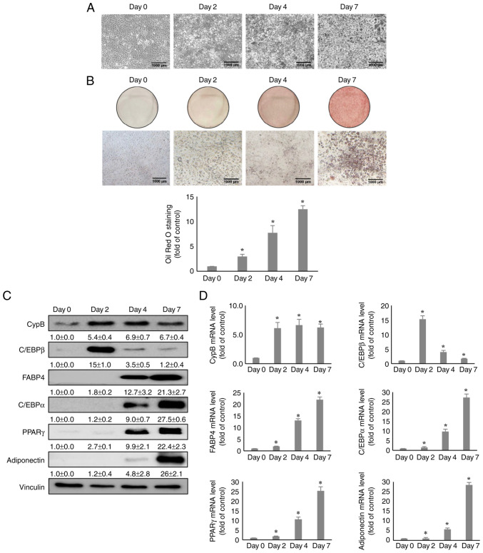
Expression of CypB during 3T3-L1 cell adipogenesis. (A) The morphology images of 3T3-L1 cells during the adipogenesis period. (B) During adipogenesis, cells (Day 0, 2, 4 and 7) were stained with lipid droplets through Oil Red O staining assay. Quantification of the stained lipid droplets was performed by measuring the absorbance at 510 nm. (C) At 0, 2, 4 and 7 days after induction of 3T3-L1 cell adipogenesis, protein levels of CypB and adipogenic markers (C/EBPβ, PPARγ, C/EBPα, FABP4 and adiponectin) during 3T3-L1 cell adipogenesis was measured by western blotting. Vinculin was used as a loading control. (D) The mRNA levels of CypB and adipogenic marker genes (C/EBPβ, PPARγ, C/EBPα, FABP4 and adiponectin) were measured by reverse transcription-quantitative PCR. The values were shown as the mean ± standard deviation of three independent experiments. *P<0.001 vs. the data of Day 0. CypB, cyclophilin B; C/EBP, CCAAT-enhancer binding protein; PPARγ, peroxisome proliferator-activated receptor γ; FABP4, fatty acid binding protein 4.

