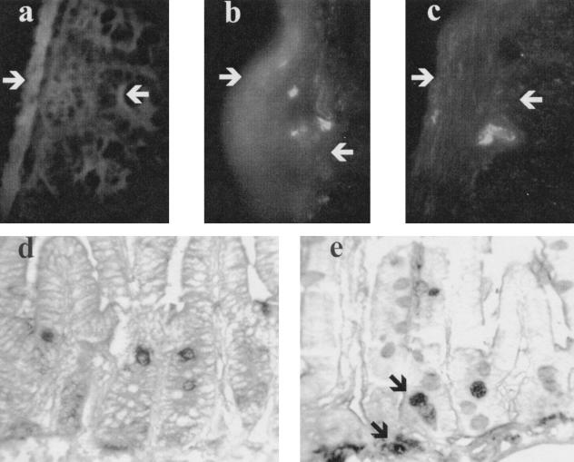
FIG. 6.
Sections of small bowel from SCID recipients 48 h after injection of CFSE-labeled spleen cells from DO11.10 RAG−/− Tg mice. The mucosa of uninfected (control) recipients does not contain CFSE-labeled cells (a), while portions of labeled cells are seen in the submucosa of C. parvum-infected recipients (b and c). White arrows mark the serosal surface and the base of the crypts. (d and e) Paraffin-embedded sections from uninfected (d) and infected (e) mice injected with BUdR 36 h after cell transfer and harvested 48 h after transfer. In panel d, only dividing epithelial cells in the crypts are labeled, while the BUdR staining of gut from infected animals shows an additional population of labeled mononuclear cells beneath the crypts (black arrows in panel e). These labeled cells have the same location as the CSFE-labeled cells identified by fluorescence microscopy.

