FIG. 1.
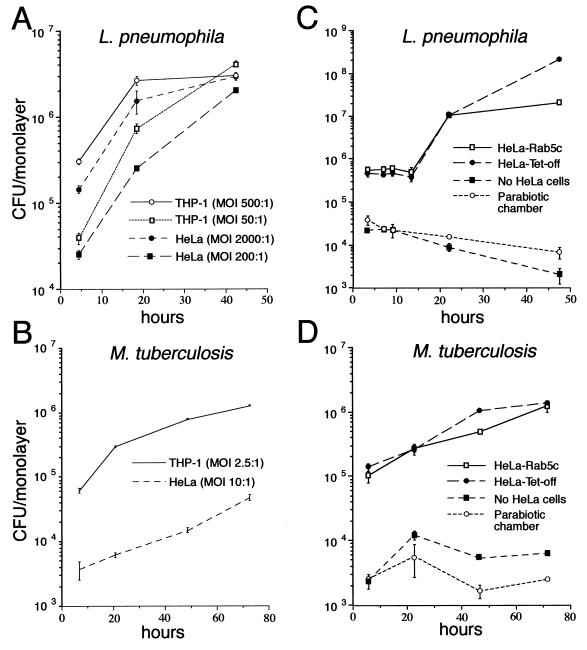
Growth of L. pneumophila and M. tuberculosis in THP-1, HeLa Tet-off, and HeLa-Rab5c cells. Monolayers of THP-1 macrophage-like cells, HeLa Tet-off cells, and HeLa-Rab5c cells expressing Rab5c were coincubated with L. pneumophila at a high MOI (2 × 10⁸/ml) or a low MOI (2 × 10⁷/ml) for 1 h (A), with L. pneumophila (2 × 10⁸/ml) for 2 h (C), with M. tuberculosis (106/ml) for 2 h (B), or with M. tuberculosis (107/ml) for 2 h (D) at 37°C, washed, and incubated in fresh medium at 37°C. At sequential times thereafter, the monolayers were lysed and combined with the culture supernatant, and the number of CFU was determined by plating serial dilutions on CYE (A and C) or 7H11 (B and D) agar plates. The capacity of the bacteria to grow extracellularly in the culture medium was assessed by inoculating L. pneumophila (C) or M. tuberculosis (D) into wells containing only the culture medium or into parabiotic chambers in which the bacteria were separated from the HeLa cells by a 0.2-μm-pore-size filter. Although the bacteria are taken up much less efficiently by HeLa cells, they multiply, once inside, with a similar doubling time in HeLa cells and THP-1 cells (A and B). Overexpression of Rab5c does not alter the intracellular growth rate of L. pneumophila or M. tuberculosis in HeLa cells (C and D). The bacteria do not grow in the absence of cell monolayers or when separated from the monolayer in a parabiotic chamber (C and D). Data shown are the means ± the standard deviations of triplicate determinations.

