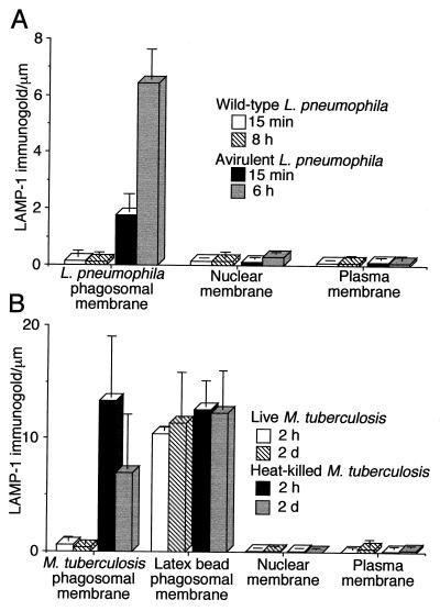
FIG. 3.
Quantitation of LAMP-1 immunogold staining in HeLa-Rab5c cells infected with L. pneumophila or M. tuberculosis. (A) HeLa-Rab5c cells were coincubated with wild-type or avirulent L. pneumophila for 15 min at 37°C and fixed immediately or coincubated at 37°C for 30 min, washed, and incubated for 6 or 8 h and then fixed. (B) HeLa-Rab5c cells expressing Rab5c were coincubated with latex beads and either live or heat-killed M. tuberculosis for 2 h and either fixed immediately or washed, incubated for 2 days at 37°C, and then fixed. After fixation, all cells were processed for cryoimmunoelectron microscopy and stained for LAMP-1. LAMP-1-bound immunogold particles were enumerated on phagosomal, nuclear, and plasma membranes. Data shown represent the mean and standard deviation of gold particle counts on at least 20 cells (each with at least one phagosome) on each of at least three electron microscopy grids. Wild-type L. pneumophila lacks LAMP-1 at both 15 min and 8 h (A). In contrast, avirulent L. pneumophila phagosomes have a modest level of LAMP-1 at 15 min and stain intensely for LAMP-1 at 6 h. Phagosomes containing live M. tuberculosis have very little LAMP-1, whereas phagosomes containing heat-killed M. tuberculosis and latex beads stain intensely for LAMP-1 at both 2 h and 2 days (B). The nuclear membrane and plasma membrane have negligible staining for LAMP-1 and serve as internal negative controls (A and B).

