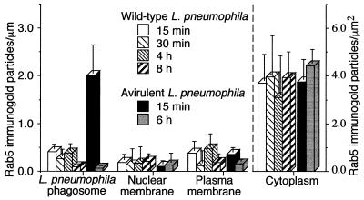
FIG. 4.
Quantitation of Rab5c immunogold staining in HeLa-Rab5c cells infected with wild-type or avirulent L. pneumophila. HeLa-Rab5c cells were coincubated with wild-type or avirulent L. pneumophila for 15 or 30 min and either fixed immediately or washed extensively, incubated for an additional 30 min to 8 h, and then fixed. After fixation, all cells were processed for cryoimmunoelectron microscopy, and Rab5c immunogold particles were enumerated on phagosomal, nuclear, and plasma membranes. Data shown are the means and standard deviations of gold counts on at least 20 cells (each with at least one phagosome) on each of at least three electron microscopy grids. (Left) At 15 min, Rab5c is scarce on wild-type L. pneumophila phagosomes but present on phagosomes containing avirulent L. pneumophila. Subsequently, Rab5c is absent or scarce on wild-type and avirulent L. pneumophila phagosomes. Rab5c is scarce on nuclear membranes and plasma membranes at all time points examined. (Right) As a control, Rab5c staining in the cytoplasm of the HeLa cells was quantitated and found to be comparable in the cells containing wild-type or avirulent L. pneumophila at all time points.

