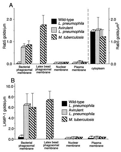
FIG. 9.
Quantitation of Rab5c and LAMP-1 immunogold staining in HeLa-Rab5c Q79L cells infected with L. pneumophila or M. tuberculosis. Monolayers of HeLa cells expressing Rab5c Q79L were coincubated with L. pneumophila or with M. tuberculosis and latex beads, fixed, and processed for immunoelectron microscopy after 30-min or 2-h incubations (respectively). The number of Rab5c-bound (A) and LAMP-1-bound (B) immunogold particles was enumerated on phagosomes, plasma membranes, and nuclear membranes. In the case of the M. tuberculosis-infected cells, vacuoles that contained only latex beads were scored as latex bead phagosomes. Vacuoles that contained both latex beads and M. tuberculosis were scored as M. tuberculosis phagosomes. Latex bead phagosomes were not scored for the L. pneumophila infected cells, due to inadequate uptake in the 30 min of coincubation. Data shown are the means and standard deviations of gold counts on at least 20 cells (each with at least one phagosome) on each of at least two electron microscopy grids.

