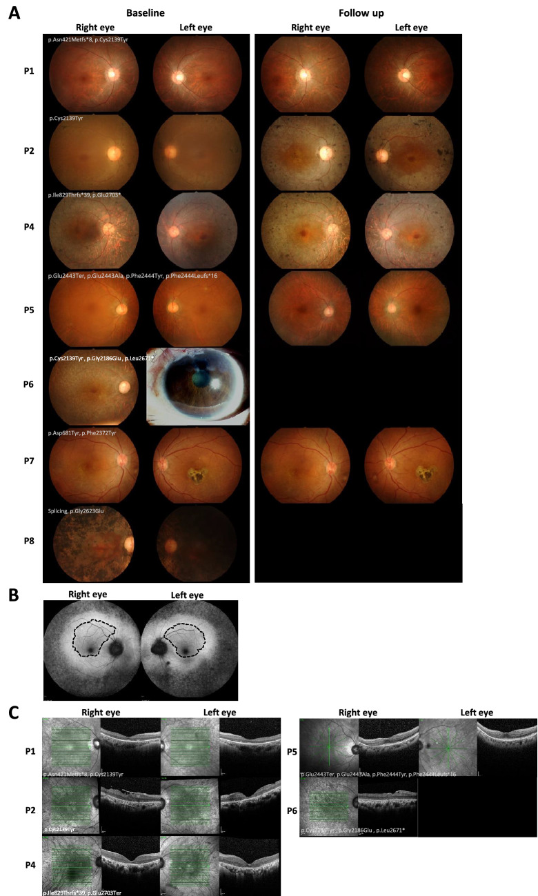
Figure 3.
Fundus photography and optical coherence tomography (OCT) of patients with EYS gene variants. (A) Fundus photographs taken from patients at baseline and at last follow up displaying retinitis pigmentosa (RP) disease progression (except for P6 and P8). (B) Fundus autofluorescence of patient 5 showed a hyperautofluorescent ring along the outer border of the macula (dashed line), with a well-preserved central foveal structure. (C) OCT of five patients at follow up.

