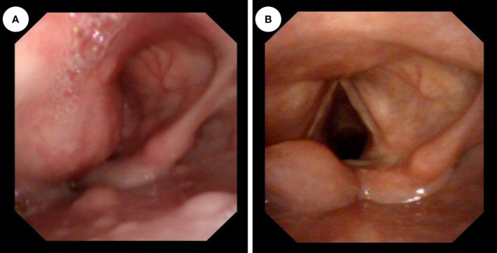
Figure 2.
(A) Preoperative fiberoptic laryngoscopy showed a smooth bulge in the left aryepiglottic fold, protruding into the laryngeal cavity, and the glottic fissure was invisible. (B) After the operation, normal laryngeal structures were observed using a fiberoptic laryngoscope, the glottic region was fully exposed, and there were no obvious abnormalities in the hypopharynx.

