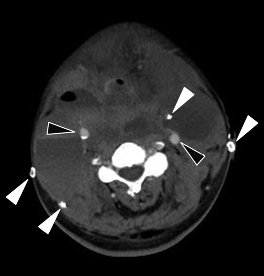
Fig. 2.

Contrast enhanced computed tomography of the neck hematoma
Axial contrast enhanced computed tomography of the neck showing a large hematoma in the bilateral neck. Although bilateral common carotid arteries (black arrows) and drain tubes (white arrows) can be identified, the preserved bilateral IJVs cannot be identified at this level.
