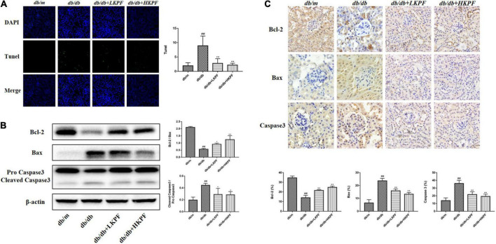
FIGURE 4.
Kaempferol reduced apoptosis in the kidneys of db/db mice. (A) TUNEL-positive cells (green) were stained in glomeruli after Kaempferol treatment, DAPI (blue) was used to stain the cellular nucleus (400×, n = 3). (B) WB assay and quantitative analysis of apoptosis-related proteins Bcl-2, Bax, and Caspase 3 expressions after Kaempferol treatment, β-actin was used as internal references; data from each group were expressed as the mean ± SD (n = 3) from three repeated WB experiments. (C) IHC assay and quantitative analysis of apoptosis-related proteins: Bcl-2, Bax, and Caspase 3 (400×, n = 5). (##p < 0.01 vs. db/m mice; *p < 0.05 and **p < 0.01 vs. db/db mice). IHC, Immunohistochemical; WB, Western blot.

