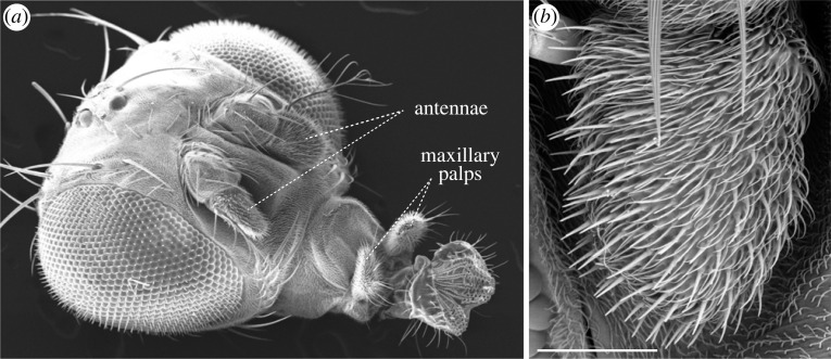
Figure 1.
Olfactory organs. (a) Scanning electron micrograph (SEM) of the head of adult D. melanogaster, showing the two bilaterally symmetric olfactory organs. Adapted from [22] (copyright © Cold Spring Harbor Laboratory Press). (b) SEM of a D. melanogaster antenna, illustrating the dense array of morphologically diverse sensilla (which house olfactory sensory neuron dendrites) covering the surface. Scale bar, 50 µm. Adapted from [23].

