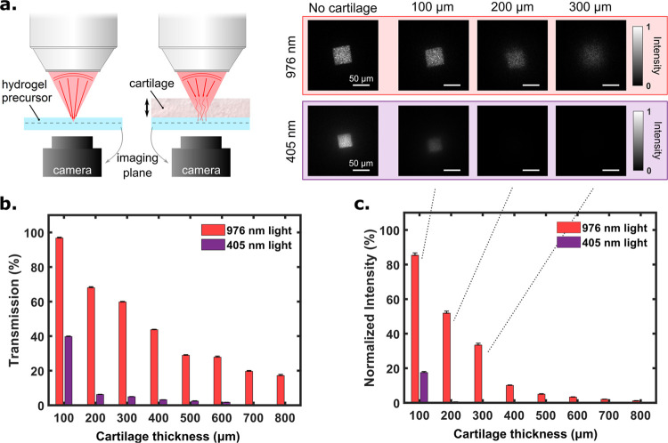
Figure 3.
Comparison between 976 and 405 nm wavelength light for penetration depth through the bovine knee cartilage. (a, left hand side) Schematic view of the experimental setup. Light (blue or NIR), at the output of a square optical fiber, is focused through the tissue on the hydrogel (containing 15 wt % gelatin solution) with a microscope objective (magnification: 20×, numerical aperture: 0.4, working distance: 20 mm). (a, right hand side) Corresponding images taken with the camera for different tissue thicknesses at the focal plane of the microscope objective. From these measurements, both (b) transmission and (c) normalized intensity (normalized by “No Tissue”) are plotted as a function of the tissue thickness.

