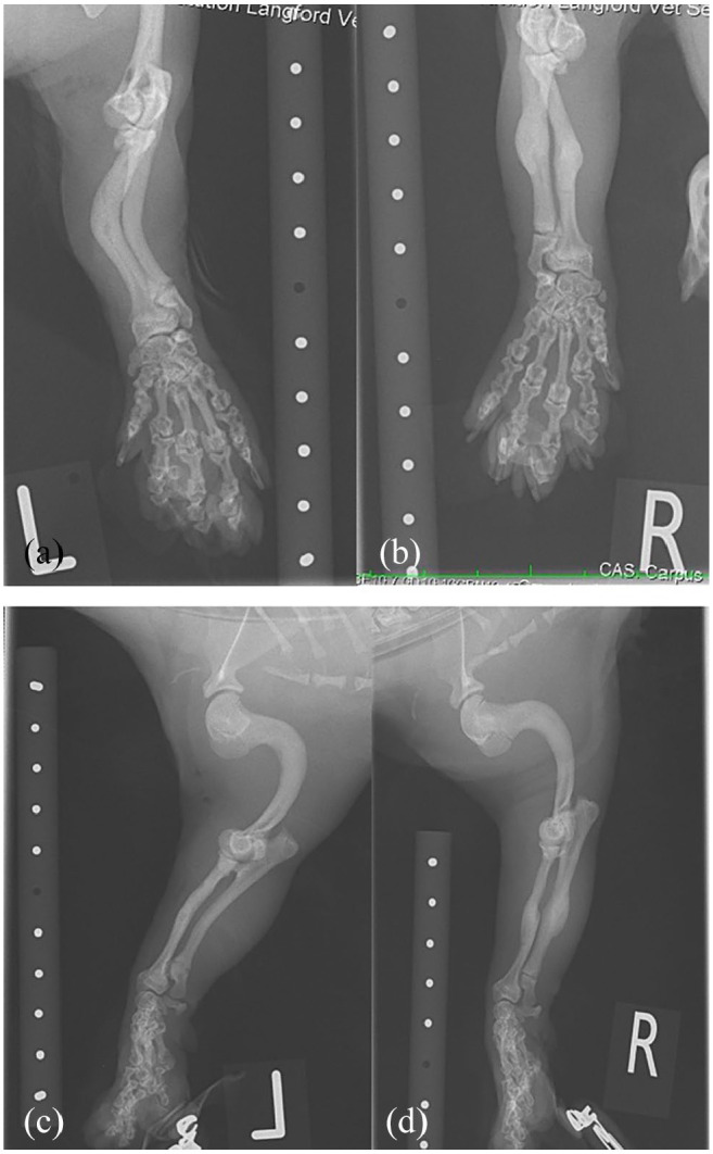
Figure 2.

Radiographs of the forelimbs of a cat with suspected pyknodysostosis and cathepsin K mutation. (a) Left and (b) right forelimb antebrachial images show mild cranial and moderate lateral curvature and thickening of the mid-diaphysis of the radius and ulna with a reduction in the normal medullary cavity. The distal radius and ulna physes appear open. The metacarpal bones and phalanges appear abnormal with very short diaphyses, more angular and wide flared epiphyses. (c,d) The humeri are markedly abnormal in shape with severe caudal curvature of the entire diaphysis, with increased medullary opacity similar to the cortical opacity
