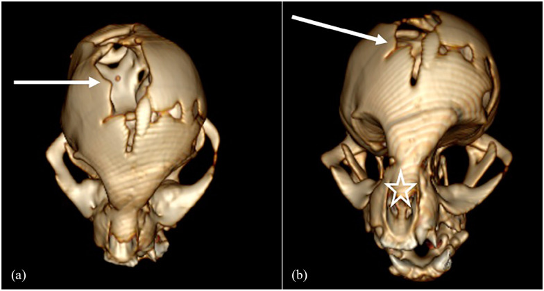
Figure 5.
Three-dimensional CT reconstruction of the skull of a cat with suspected pyknodysostosis and cathepsin K mutation. (a) Dorsal plane of the cat’s skull shows a large defect at the level of the fontanelle between the frontal and parietal bones (arrow). (b) Dorsal plane of the cat’s skull shows a large defect at the level of the fontanelle (arrow) and marked deviation of the nasal septum (star)

