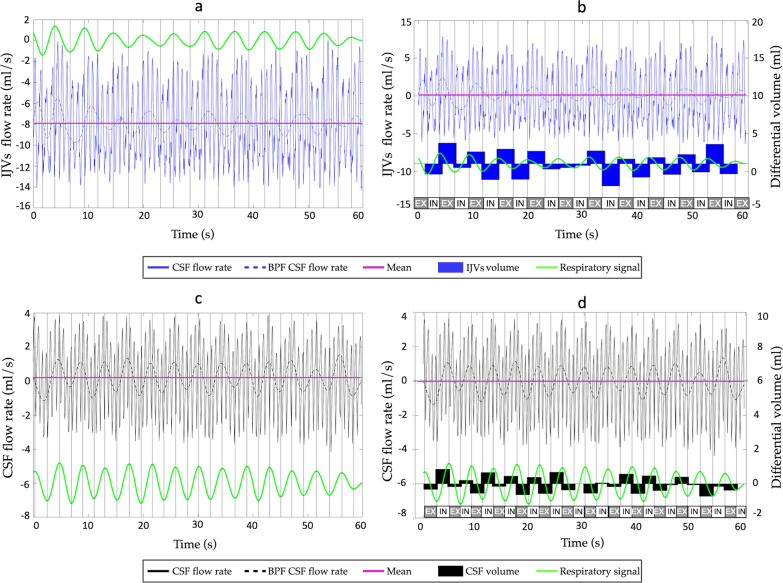
Fig. 3.
Differential flow volume (in ml) computed during each inspiration and expiration. Respiratory signal (green), flow rate curve and its mean value (purple) are shown in (a) and (c) for the Internal Jugular Veins (IJVs—blue curve) and cerebrospinal fluid (CSF—black curve) respectively. In b and d, the mean-centered flow rates are shown (flow rate curves minus its mean value) for the IJVs (b) and CSF (d). The inspiration (in) and expiration (ex) phases are written, based on the respiratory signal: its rising parts were considered inspirations. The differential volumes (integrals of the mean-centered flow rate over the inspiratory and expiratory phases) are shown as black bars in (b) for the IJVs and (d) for the CSF

