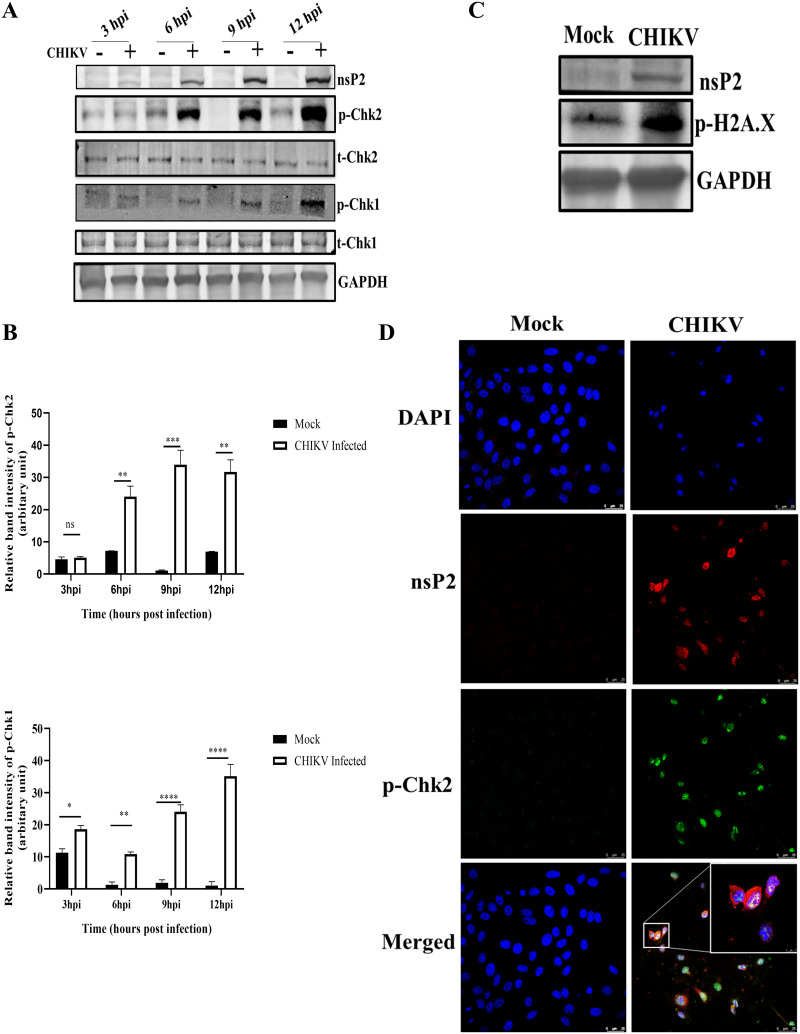
FIG 1.
CHIKV infection induces DDR pathways. The Vero cells were mock or CHIKV infected and harvested at various time points. (A) Western blotting was performed using the nsP2, p-Chk2 (Thr-68), total Chk2, p-Chk1(Ser354), total Chk1, and GAPDH antibodies. (B) Bar diagrams showing relative band intensities of p-Chk2 and p-Chk1 at different time postinfection. Data of three independent experiments are shown as mean ± SD. (C) Western blot depicting the level of p-H2A.X (Ser-139) in mock and infected samples. (D) Mock or infected Vero cells were stained with the p-Chk2 and nsP2 antibodies. Nuclei were counterstained with DAPI. Scale bar = 25 μm. *, P ≤ 0.05; **, P ≤ 0.01; ***, P ≤ 0.001; and ****, P ≤ 0.0001 were considered statistically significant. ns, not significant.

