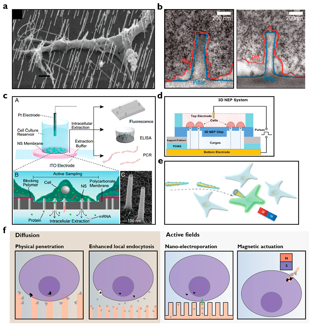
Figure 2.

Modes of intracellular cargo delivery using nanostructures. (a) Electron microscopic image showing penetration of neuron by nanowires25 (scale bar, 1 μm) in contrast to the images in panel b where neurons were found wrapping their membrane around nanopillars.12 (c) The nanostraw electroporation platform where cultured cells on the platform are exposed to local electric fields for controlled nanoelectroporation for delivery and extraction of materials such as mRNA and proteins into and out of the cells.16 Electron microscopic image shows nanostraws of about 1.5 μm in height and 150 nm in diameter. (d) Nanochannel electroporator capable of high throughput (40[IPS] 000 cells/cm2) delivery of cargo into cells.15 (e) Magnetic nanospears functionalized with plasmid encoding for green fluorescent protein (GFP). A benchtop magnet can be used to guide the nanospears for targeted single cell transfection.17 (f) Proposed mechanisms of intracellular cargo delivery using nanostructures. Cargo delivery is likely to occur through both physical penetrations followed by passive diffusion and cargo diffusion followed by endocytosis. By coupling of active fields, intracellular cargo delivery can also occur through nanoelectroporation and magnetic actuation. Note that the illustration is for nonadherent or suspension cells. Adherent cells typically conform to the shapes of the nanostructures after an hour of culture. Images adapted with permission from the following: ref 12, Copyright 2012 American Chemical Society; ref 15, Copyright 2016 Royal Society of Chemistry; ref 16, Copyright 2017 Proceedings of the National Academy of Sciences; ref 17, Copyright 2018 American Chemical Society; ref 25, Copyright 2007 American Chemical Society.
