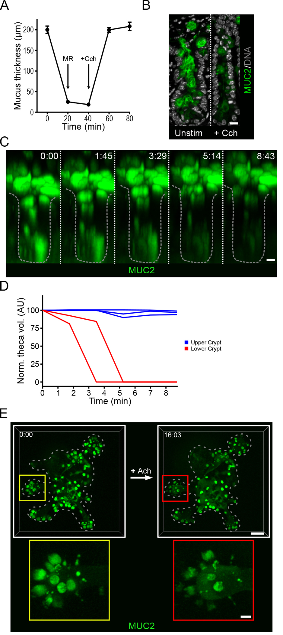
Fig. 1. Cholinergic stimulation of the small intestine induces a crypt-specific secretory response.

(A) Mucus thickness in mouse ileum immediately after explanation (time 0), after mucus removal (MR) at 20 min, and at the indicated time points after the tissue was stimulated by basal perfusion with carbachol (Cch) at 40 minutes. Data presented as mean ± SD (n = 5 independent experiments). (B) Immunofluorescence staining for Muc2 in goblet cells in the crypts of unstimulated (Unstim) and Cch-stimulated ileal explant tissue. Scale bar, 10 μm. Image is representative of 3 independent experiments. (C) Multi-photon live tissue imaging of small intestinal crypts from RedMUC298tr mice) after the tissue was stimulated with Cch at time 0. Scale bar, 10 μm. Images are representative of n = 3 independent experiments. (D) Quantification of theca volume of individual mCherry+ cells from the crypt in panel C. (E) Live confocal imaging of ileal organoids generated from RedMUC298tr mice and stimulated with acetylcholine (Ach). Scale bar, 50 μm (main images) and 10 μm (insets). Images are representative of n = 3 independent experiments.
