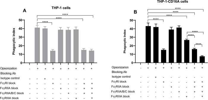
Fig 5. FcγR utilization by THP-1 and THP-1-CD16A cells in the phagocytosis of IgG-opsonized human erythrocytes.
THP-1 cells were differentiated to macrophages by treatment with PMA (100 ng/mL) as described in the methods. (A) THP-1 cells. (B) THP1-CD16A cells. Opsonization: (-) indicates erythrocytes were non-opsonized (incubated with phosphate buffered saline), (+) indicates erythrocytes were opsonized with a polyclonal anti-human RhD antibody (WinRho SDFTM). The contribution of each FcγR to the phagocytosis was evaluated using Fc region deglycosylated blocking antibodies (final concentration of 10 μg/mL each): anti-FcγRI (clone 10.1), anti-FcγRIIA (clone IV.3), anti-FcγRIIA/B/C (clone AT10), or anti-FcγRIIIA (clone 3G8). The deglycosylated mouse IgG1 (clone MOPC-21) and deglycosylated mouse IgG2b (clone MPC-11) were used in combination as isotype controls (final concentration of 10 μg/mL each). The phagocytic index was calculated as the number of erythrocytes engulfed per 100 macrophages. Data are presented as the mean ± the standard deviation of five independent experiments. For panels A and B, the statistical analysis was performed with a one-way analysis of variance (ANOVA) and Tukey’s multiple comparisons test (****: p <0.001).

