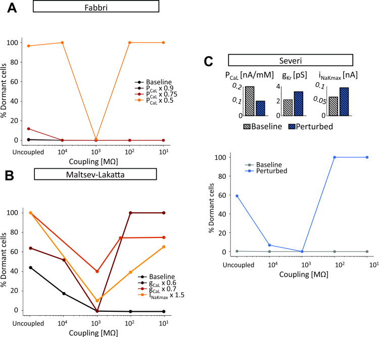
Fig 7. Pathophysiological changes in ionic currents lead to a pattern of tissue automaticity dependent on the degree of intercellular coupling.
(A) Effect of L-type Ca2+ current (ICaL) perturbation in the Fabbri tissue model (σ equal to 0.1). (B) Effect of perturbation in ICaL and Na+/K+ pump (INaK) in the Maltsev-Lakatta tissue model (σ equal to 0.4). (C) Effect of combined ICaL, rapid delayed rectifier K+ current (IKr), and INaK perturbation in the Severi tissue model (σ equal to 0.2).

