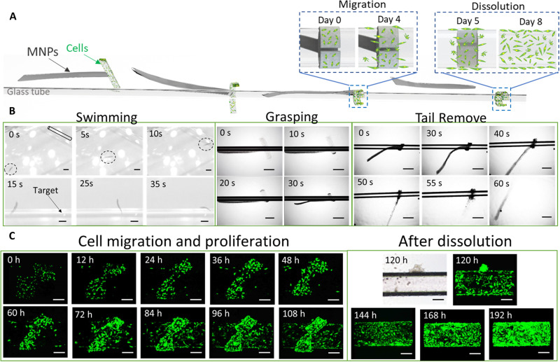
Fig. 7. Soft microrobotic cell delivery demonstration through physical contact.
(A) The MMR is moved to the target area, attached to the structure, grasped the target through the functional module, delivered the cells, and removed the propulsion module through magnetic control and transformation under ionic stimulation. (B) Optical images showing the MMR propulsion, self-trapping of the tubular target area, and removal of the magnetic module after the cell delivery was completed. Scale bars, 1 mm. (C) Fluorescent optical microscopic images of GFP-enhanced HUVEC proliferation and migration after the MMR grasped the target area; the delivered cells fully covered the target area after the removal of the propulsion module. Scale bars, 200 μm.

