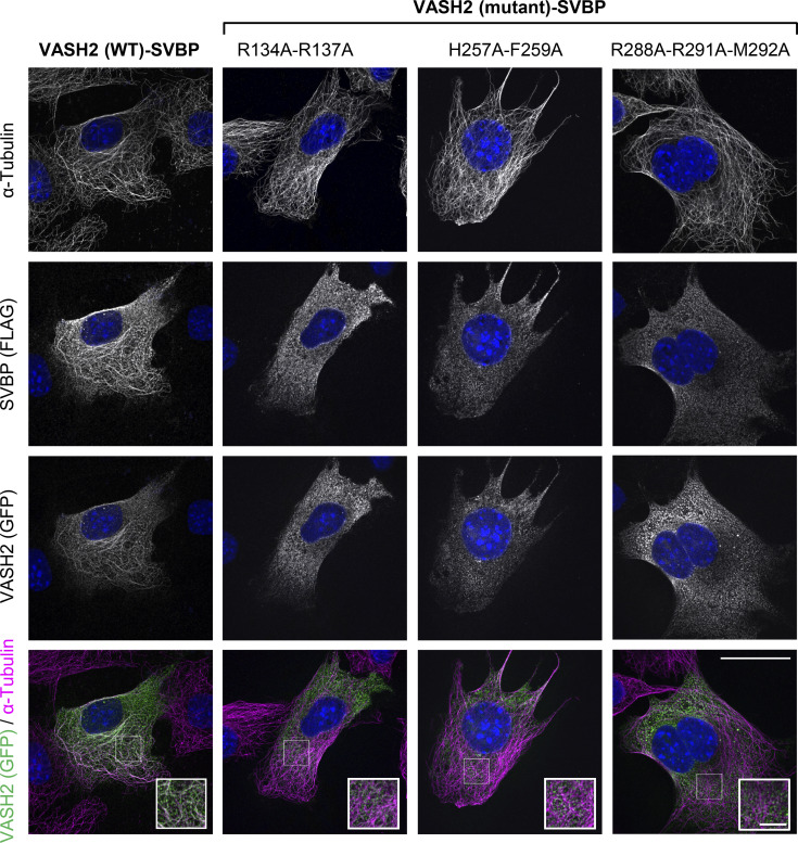
Figure S4.
VASH2–SVBP mutants associate poorly with microtubules in cells. Representative immunofluorescence images of NIH3T3 fibroblasts cotransfected with plasmids encoding WT or indicated VASH2-eGFP mutants and SVBP-myc-FLAG. Merged images of VASH2-eGFP (green) and α-tubulin (magenta) stainings are displayed in the bottom lane. Insets are magnifications of the white square regions. Scale bars, 25 µM for entire images and 5 µM for insets.

