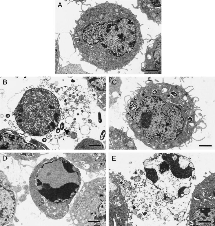
FIG. 5.
Electron micrographs of J774 cells. (A) Uninfected J774 cells as a control. (B) J774 cells 1 h after infection with CHA, showing oncotic morphology: flocculation of the chromatin, dissolution of the cytoplasm, and swollen nuclei. (C) J774 cells 1 h after infection with the CHA-D1 strain. (D and E) J774 cells treated by UV-irradiation showing either early apoptotic morphology with intense perinuclear chromatin aggregation but cytoplasm integrity (D) or late apoptotic morphology with the nucleus having the same profile as in early apoptosis but dissolution of the cytoplasm (E). The arrow indicates bacteria. Bars, 2 μm.

