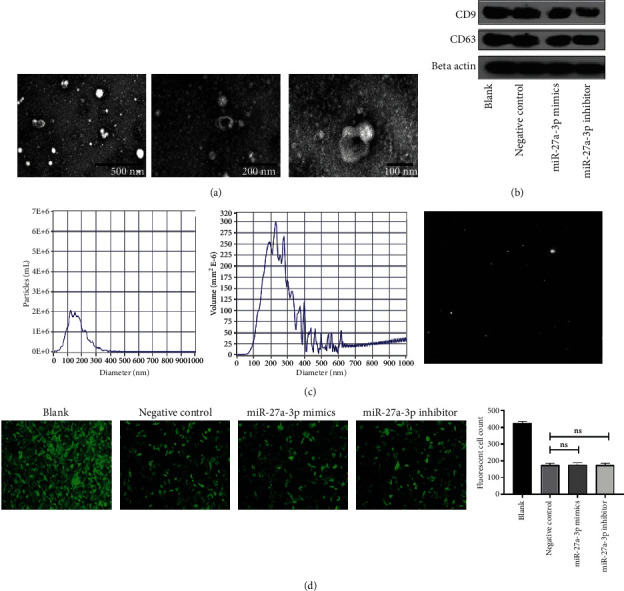
Figure 5.

Exosomes identification. (a) Transmission electron microscopy identification of exosomes. (b) Exosome markers CD9 and CD63 confirmed by western blot. (c) Exosomes volume and purity. (d) miR-27a-3p transfection efficacy indicated by fluorescence microscopy (scale bar 50 μm).
