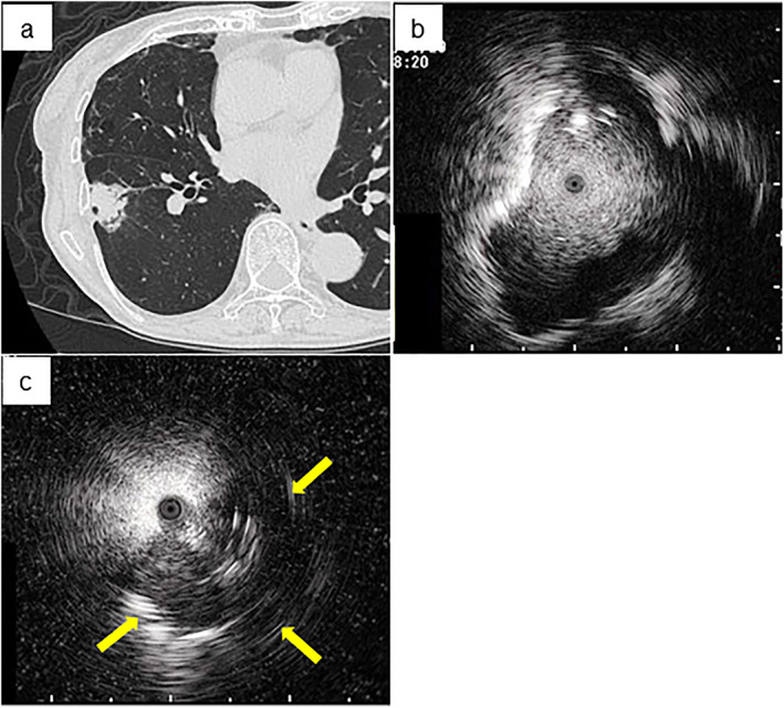
FIGURE 2.

Representative images of the bronchus sign and endobronchial ultrasonography with a guide sheath (EBUS) imaging including within and adjacent to the lesion. (a) The bronchus sign is observed in the peripheral lung field on chest computed tomography with the lung window setting. EBUS imaging of (b) within and (c) adjacent to (arrow shows the detected image of the targeted lesion) the image are shown
