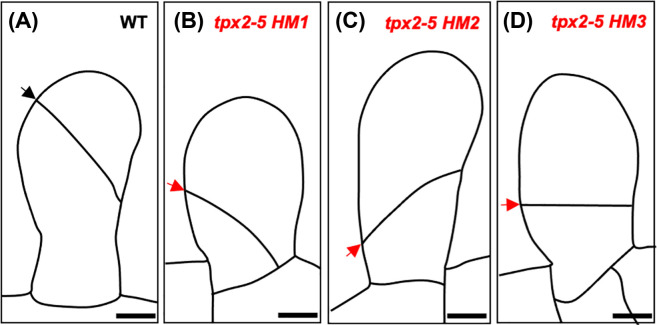
Figure 4. The hypomorphic tpx2-5 mutants.
Schematic diagrams of the first division of the gametophore apical cell in wild-type (A) and incorrectly oriented divisions of the gametophore apical cell in the hypomorphic mutants tpx2-5 HM1 (B), tpx2-5 HM2 (C) and tpx2-5 HM3 (D). Correctly and incorrectly oriented division planes are denoted by black and red arrows respectively. Scale bars represent 10 µm. Images adapted from [25].

