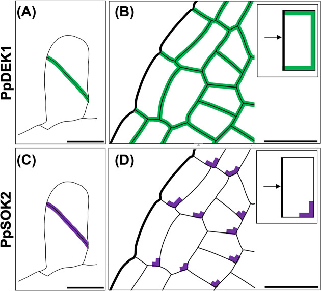
Figure 5. Polar localization of PpDEK1 and PpSOK2.
(A, B) Polar localization of PpDEK1 following the first division of the gametophore apical cell (A) and in developing phyllids (B). Inset in (B) shows a notable absence of PpDEK1 localization at phyllid margins. (C, D) Polar localization of PpSOK2 following the first division of the gametophore apical cell (C) and in developing phyllids (D). Inset in (D) highlights that PpSOK2 is localized at the base of cells within the phyllids and is enriched in the corners that are more distal to the phyllid margins. Scale bars represent 20 µm. Images adapted from [37,51].

