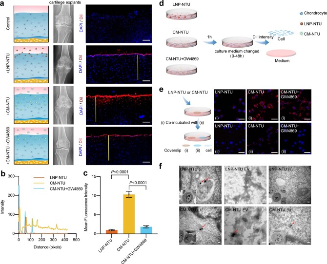
Extended Data Fig. 3. Superior tissue penetration of the CM-NTUs.
(a) Degenerated tissues and organs often exhibit fibrosis and densification of the ECM. To deliver an intra-articular injected nanomedicine to the cytosol of chondrocytes in knee joints, researchers must overcome the biological barrier for deep penetration into the avascular and dense degenerated cartilage58. Cartilage explants from OA patients were examined to assess the depth of CM-NTU or LNP-NTU (NTUs labelled with DiI) penetration after 24 h. For the inhibitor group, cartilage explants were pretreated with an extracellular vesicle (EV) secretion inhibitor for 24 h before CM-NTU incubation (scale bar, 100 μm; nuclei, blue; NTUs, red). (b) DiI fluorescence intensity profiles across the section along the selected line (indicated by a yellow line in the inset image of a). (c) Mean fluorescence intensity of cartilage sections (n = 3, mean ± SD). (d) Schematic diagram of the detection of DiI fluorescence intensity in chondrocytes and culture medium. (e) Intracellular transfer of CM-NTUs or LNP-NTUs (NTUs labelled with DiI) visualized by confocal microscopy (scale bar, 20 μm; NTUs, red; nuclei, blue). Chondrocytes on coverslips (i) were cultured in medium that contained CM-NTUs or LNP-NTUs for 6 h. A coverslip (i) was rinsed and imaged. Then, fresh culture medium was added along with a coverslip with fresh cells on the coverslip (ii) for 24 h. For the inhibitor group, chondrocytes were pretreated with an EV secretion inhibitor for 24 h. The DiI signal from the CM-NTUs was high in the cells on coverslips (ii), indicating that some of the NTUs taken up in the cells on coverslips (i) were transported into the medium and subsequently internalized by the cells on coverslips (ii). In contrast, the NTUs from the LNP-NTU-treated cells exhibited limited transportation to the cells on coverslips (ii). Moreover, pretreatment with GW4869 blocked the transcellular transmission effect of the CM-NTUs. (f) Intracellular transfer of CM-NTUs or LNP-NTUs (gold nanoparticles encapsulated into NTUs) visualized by transmission electron microscopy (TEM) (scale bar, 200 nm). The processing of chondrocytes on the coverslip is the same as that described in e. The EVs secreted by the chondrocytes on the coverslip (i) were collected separately for TEM observation. The cells stimulated by the CM-NTUs could secrete EVs containing gold nanoparticles, and gold nanoparticles could be observed in the cells on coverslips (ii). In contrast, no gold nanoparticles were observed in EVs secreted by the LNP-NTU-stimulated cells or the cells on coverslips (ii). n represents the number of human specimens. P values are indicated in the graph and were determined using one-way ANOVA (c).

