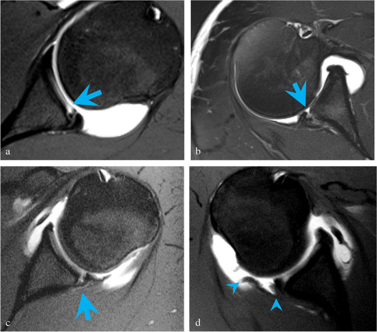
Fig. 4.
a Axial T1-weighted image with fat suppression demonstrates posterior glenolabral articular disruption (PLAD) with tear of the articular cartilage (blue arrow) in a 17-year-old male pitcher with 6 months of pain and instability following an acute subluxation event during batting. Arthroscopy showed a tear of the posterior labrum extending from 6-o’clock to 9-o’clock position with fraying of the posteroinferior glenoid cartilage. b Axial T2-weighted image with fat suppression shows a reverse Bankart lesion (blue arrow) in a 32-year-old male with pain and instability following a hockey injury. A slightly displaced bony fragment was found at surgery. The posterior labrum was noted to be attached to the bony fragment but torn away from the remaining glenoid. c Axial T1-weighted image with fat suppression shows a posterior labrocapsular periosteal sleeve avulsion (POLPSA) with stripping of the periosteum (blue arrow) in an 18-year-old male with 6 months of pain and recurrent subluxations following an acute dislocation event with self-reduction playing football. Arthroscopy confirmed a posterior labral tear extending from 7 o’clock to 9 o’clock. d Axial T1-weighted image with fat suppression demonstrates a posterior humeral avulsion of the glenohumeral ligament (PHAGL) (avulsed ligament delineated by blue arrowheads) in a 17-year-old male with 3 months of pain and recurrent subluxations following an acute posterior dislocation event while checking another player during a hockey game. Arthroscopy confirmed a tear from the 6 o’clock to 10 o’clock position. The posterior capsule and posterior band of the inferior glenohumeral ligament were found to be avulsed from the insertion on the humeral head

