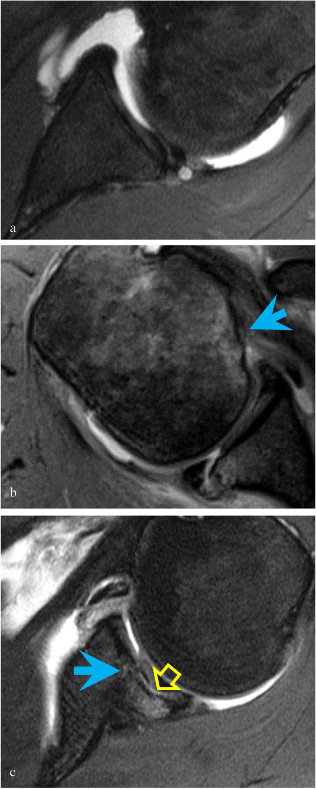
Fig. 5.

a Axial T2-weighted image with fat suppression shows a paralabral cyst in a 32-year-old male with an isolated posterior labral tear. b Axial T2-weighted image with fat suppression shows a humeral fracture (reverse Hill-Sachs) with cortical irregularity and flattening of the humeral head (blue arrow) in a 47-year-old male who suffered a posterior shoulder dislocation during a seizure and with 2 weeks of recurrent subluxations. c Axial T2-weighted image with fat suppression shows a glenoid fracture (reverse bony Bankart) with focal fracture and depression of the glenoid (blue arrow) and cartilage delamination (yellow open arrow) in a 31-year-old male former softball player who at the time of imaging unloaded heavy parcels at work. The osteochondral injury was noted to be unstable at surgery
