Figure 5.
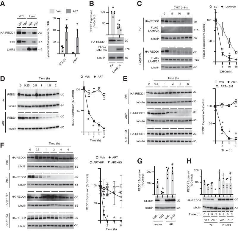
REDD1 is degraded by chaperone-mediated autophagy. A: HA-REDD1 was expressed in REDD1-KO MIO-M1 cells. Lysosomes were isolated by subcellular fractionation from cells exposed to the CMA-activator AR7 or vehicle (Veh) for 1 h. REDD1, c-fos, and LAMP2 expression were evaluated in whole-cell lysate (WCL) vs. lysosomes (Lyso) by Western blotting. Representative blots are shown. Protein molecular mass in kDa is indicated at right of blots. REDD1 and c-fos lysosomal association was quantified. B: HEK293 Tet-On HA-REDD1 cells were transfected to express FLAG-tagged LAMP2A or an empty vector (EV) control. REDD1, LAMP2A, and tubulin protein expression was evaluated by Western blotting. C: REDD1 degradation was evaluated in HEK293 Tet-On HA-REDD1 cells expressing FLAG-LAMP2A or EV by cycloheximide (CHX)-chase (n = 4). D: HEK293 cells were exposed to tunicamycin for 4 h to promote endogenous REDD1 expression and subsequently exposed to AR7 or Veh (n = 5). E: HEK293Tet-On HA-REDD1 cells were exposed to culture medium containing AR7 in the presence/absence of the autophagy inhibitor bafilomycin (n = 4). F: HA-REDD1 was expressed in REDD1-KO MIO-M1 cells by transient transfection. Cells were exposed to medium containing 30 mmol/L glucose (HG) or H2O2 (HP) for 24 h prior to addition of AR7 or Veh (n = 4). G: Endogenous REDD1 expression was evaluated in HEK293 cells pretreated with HP or water as a control for 2 h prior to exposure to AR7 or Veh for 2 h. H: WT or K129R HA-REDD1 variants were expressed in REDD1-KO MIO-M1 cells by transient transfection (n = 4). Cells were exposed to Veh or AR7 as indicated. Data are represented as mean ± SD. *P < 0.05 vs. Veh or EV; #P < 0.05 vs. water or WT.

