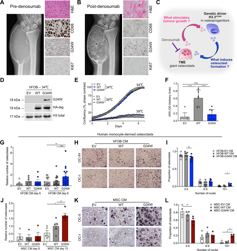
Fig. 1. Mutant H3.3G34W in osteoprogenitors regulates bone formation and stimulates osteoclast recruitment.
A, B GCT of the distal ulna in a 35-year-old male. A Pre-denosumab treatment: anteroposterior radiograph of the distal ulna shows the tumour (dotted outline) without mineralisation. H&E-stained section: features of a benign GCT with CD68-positive-osteoclasts interspersed with proliferative H3.3G34W-mononuclear stromal cells (G34W; Ki67). B Post-denosumab treatment: after one month’s treatment showing similar tumour size (dotted outline) to A but prominent mineralisation (asterisk). H&E-section: bone formation (asterisk) and absence of CD68-positive osteoclasts but persistent H3.3G34W-neoplastic cells (G34W) with a reduced proliferative index (Ki67) compared to A. C Schema of proposed interactions between stromal/osteoprogenitors and TME. H3.3G34W-osteoprogenitor-derived factor(s) result in an environment permissive for formation of abnormally large osteoclasts, which secrete factor(s) stimulating tumour growth. Denosumab treatment results in depletion of osteoclasts and removes the growth stimulus for stromal/osteoprogenitors. D Representative western blot of HA-tagged-H3.3 and H3.3-G34W expression in transfected hFOBs. E Proliferation of undifferentiated hFOBs grown at 34 °C and differentiated at 39 °C for 6 days in mineralisation medium assessed by Incucyte; 2 experiments, 3 replicates per experiment. F Quantification of ARS mineralisation of hFOBs on day 6 of differentiation. 8 replicates, three experiments. G, J Number of osteoclasts generated in the presence of conditioned medium (CM) from (G) undifferentiated (day 0, 34 °C) and differentiated (at 39 °C for 6 days) hFOB (number relative to WT CM day 6) and (J) from iPSC-derived-MSCs differentiated to osteoblasts for 8 and 15 days (number relative to WT CM day 15). 7–18 (G) and 4-5 (J) osteoclast preparations. H, K Representative tartrate-resistant acid phosphatase (TRAP) staining of two osteoclast cultures (OC-I and II) in the presence of CM from (H) hFOBs differentiated for 6 days or (K) iPSC-derived-MSCs differentiated to osteoblasts for 15 days; 8 osteoclast preparations. 4× magnification. I, L Quantification of number of nuclei per osteoclast in H and K expressed as proportion of OCs, calculated as the number of OCs exhibiting 3–5, 6–9 or ≥10 nuclei over the total number of OCs; results from 8 (I) and 4 (L) osteoclast preparations. Data are mean ± SD (F), ±SEM (G, J, I, L). F 1-way ANOVA. G, J: 1-way repeated measures (RM)ANOVA for each time point. I, L: 2-way RM ANOVA.

