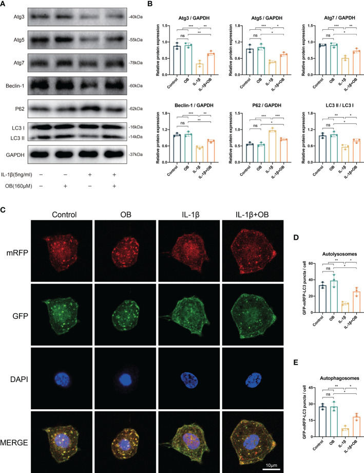
Figure 6.
OB rescued the impaired autophagy process in IL-1β–induced chondrocytes. Chondrocytes were administered with IL-1β (5 ng/ml) or/and OB (160 μM) for 24 h. (A) Western blot and (B) quantitative analysis showed the impaired autophagy induced by IL-1β could be rescued by the treatment of OB (160 μM). (C) Chondrocytes were transfected with tandem GFP-RFP-LC3 adenovirus, and the strength of autophagic flux was captured with a confocal microscope. (D, E) Quantitative analysis of autolysomes (red puncta) and autophagosomes (yellow puncta) among groups. Data were presented as means ± SD (n = 3). ns, no significance; *p < 0.05; **p < 0.01; ***p < 0.001.

