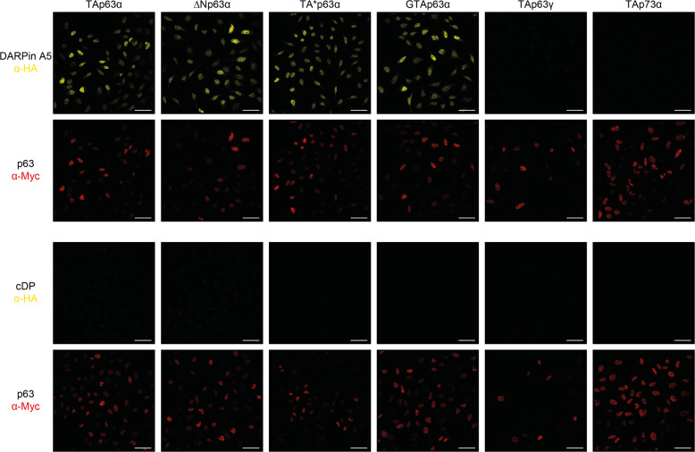
Fig. 5. Detection of different p63 isoforms in stably expressing HeLa cells.
Cells expressing the indicated p63 isoforms or as a control TAp73α, were fixed with formaldehyde and incubated with HA-tagged DARPin A5, followed by the goat anti-HA antibody (a190138a - Bethyl) and the secondary antibody Alexa Fluor 568 anti-goat (A11057—Life Technologies). The same cells were also stained with the mouse anti-myc antibody 4A6 (Millipore) and the secondary antibody Alexa Fluor 647 anti-mouse (A31571—Life Technologies) as all p63 isoforms and TAp73α are labeled with an N-terminal myc-tag. All SAM domain-containing p63 isoforms show strong staining while TAp63γ which lacks a SAM domain does not show any signal above background. Cells expressing TAp73α, which has a SAM domain, do not show staining either demonstrating the specificity of the DARPin A5 for the p63 SAM domain. A control DARPin does not show any signal above background. Scale bar, 50 µm.

