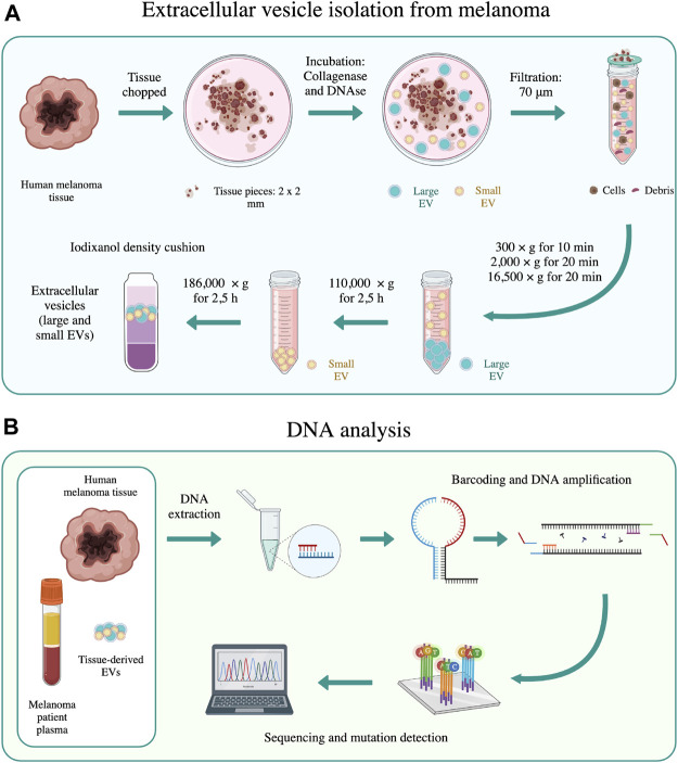
FIGURE 1.
Schematic representation of the main methods used in this work. (A) EV isolation from melanoma tissues was performed according to the method of Crescitelli et al. (Crescitelli et al., 2021). (B) DNA sequencing was performed on human melanoma tissue, tissue-derived EVs, and melanoma patient plasma using SiMSen-Seq analysis, as described by Ståhlberg et al. (Ståhlberg et al., 2017). The schematic was created with biorender.com.

