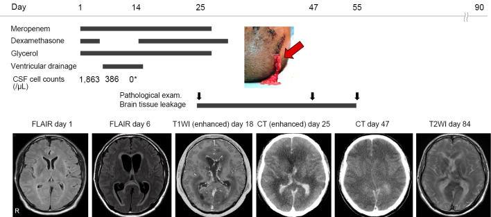
Figure 1.
Clinical course. A patient with Listeria monocytogenes meningitis was initially treated with meropenem and dexamethasone. Although the CSF cell count decreased after antibiotic treatment, brain MRI revealed hydrocephalus with ventriculitis and diffuse cerebral edema on day 6. He underwent external ventricular drainage but was ventilated due to worsening cerebral edema that persisted for two months. On day 25, brain tissue had leaked through the incision site of the external drain (red arrow) due to the severe cerebral edema lasting over a month. While cerebral edema persisted, the pathology of the leaked specimen showed significant neutrophil infiltration. On day 55, the brain tissue stopped leaking, indicating that the cerebral edema had finally subsided. A specimen revealed no neutrophils. *The CSF cell count (0 cells/μL) shown at hospital day 14 was corrected by the red blood cell count. CSF: cerebrospinal fluid, MRI: magnetic resonance imaging, FLAIR: fluid-attenuated inversion-recovery, T1WI: T1-weighted imaging, T2WI: T2-weighted imaging, CT: computed tomography

