Figure 7.
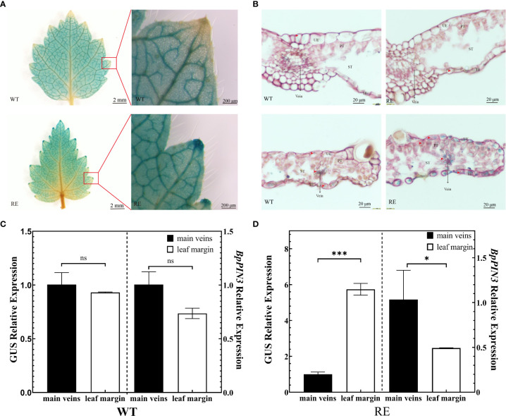
Asymmetric distribution of auxin in the leaves from the RE lines (A) GUS staining of the leaves from the RE and WT lines, Scale bar = 2 mm and enlarged at the leaf margin, Scale bar = 200 μm; (B) Slice observation after GUS staining in the main vein region and leaf margin region, Scale bar = 20 μm; (C) GUS and BpPIN3 expressions in the WT lines; (D) GUS and BpPIN3 expressions in the RE lines. Error bars indicate SD, n = 3. Data were analyzed by Tukey’s tests. *p< 0.05; ***p< 0.001. ns, not significantly different.

