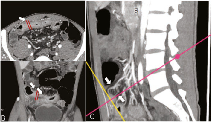
FIGURE 3.

Images from a male patient with Crohn's disease. Transverse (a), coronal (b), and sagittal (c) enhanced CT images showed bowel wall thickening and luminal narrowing of the jejunum (arrow); the MCFI of the designated segment reconstructed from the adjacent mesenteric vessels was scored with 2, because a quarter of the bowel surface was covered by the mesenteric vessels. Note: The MCFI, which reflects the degree of mesenteric fat wrapping around the gut, is scored from 1 to 8 according to the areas of bowel surface covered by the corresponding mesenteric vessels. CT, computed tomography; MCFI, mesenteric creeping fat index.
