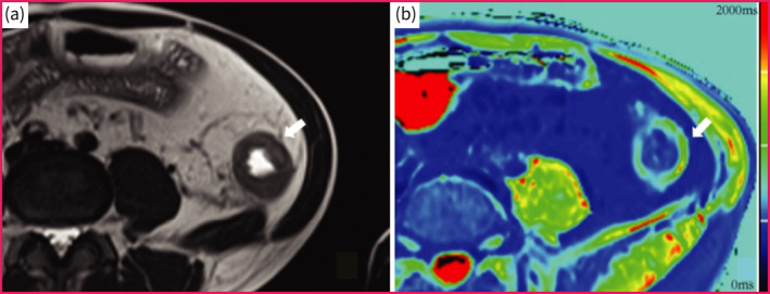
FIGURE 4.

T1 mapping of a f Crohn's disease suffered female patient with moderate‐to‐severe fibrosis in descending colon. (a) Axial T2‐weigheted image showed bowel wall thickening of the descending colon. (b) The T1 mapping shows that the T1 value of affected descending colon was 1430 ms.
