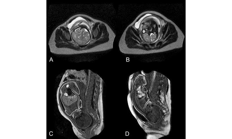
Figure 3.

(A) T2 gradient echo sequence showing an axial slice centered on the fetal brain at the superior inlet level. (B) The same sequence at the iliac bone level. (C) T1 gradient echo sequence in the sagittal plane showing bony pelvis and fetal head before entering into labor. The empty bladder is in retropubic position. (D) Same T1 gradient echo sequence showing the fetal head molding in the middle brim during second phase of labor with a full bladder. The fetal head is rotated in occipito-pubic position, and the full bladder is ascended above the upper limit of the pubic bone.
